lv segments on echo|lv segments diagram : 2024-10-22 Complete review on left ventricular systolic function, with emphasis on echocardiography, definitions, methods and guidelines. Eyedro Business 3-Phase Energy & Solar Monitor | Net Metering | High-Resolution Electricity Usage Data via My.Eyedro.com -No Fee | Alerts | Reports | Real-time Energy Costs | E5B-EW-E3 (Ethernet/WiFi) Visit the Eyedro Store. 4.2 28 ratings. $34900. Style: Ethernet/WiFi (E5B-EW-E3)
0 · what is fractional shortening echo
1 · wall motion chart echo
2 · lv wall motion abnormalities
3 · lv segments diagram
4 · lv function assessment by echo
5 · how to assess lv function
6 · 17 segments of the heart
7 · 17 segments of left ventricle
In this asymptomatic patient, with a 3-dimensional left ventricular ejection fraction (LVEF) of 58% (left), the mean global longitudinal strain (GLS) was 14%, showing both spatial and temporal variation in the 4-, 2-, and 3-chamber views.
lv segments on echo*******Although certain variability exists in the coronary artery blood supply to myocardial segments, segments are usually attributed to the three major coronary arteries. Visual .Herein we review the conventional assessment of LV systolic function and examine the role of speckle-tracking echocardiography (STE), a new method to assess LV function. We also highlight the role of STE in the .
tricular [LV] size and ejection fraction [EF], left atrial [LA] volume), outcomes data are lacking for many other parameters. Unfortunately, this approach also has limitations.
Complete review on left ventricular systolic function, with emphasis on echocardiography, definitions, methods and guidelines.A normal LV ejection fraction in the presence of the heart failure syndrome leads to a search for diastolic dysfunction. Typical echo findings in diastolic dysfunction are normal LV cavity size, thickened ventricle, and reversed . LV wall has been divided into 17 segments for the ease of the assessment of regional function. Apical segment, also called apical cap, is the only segment without a .Strain echocardiography, performed by using the speckle-tracking technique, can identify subclinical left ventricular dysfunction before left ventricular ejection fraction declines.lv segments diagramThis chapter demonstrates left chamber quantification through various measurements of left ventricular size and dimensions, left ventricular mass, left ventricularglobal function, .
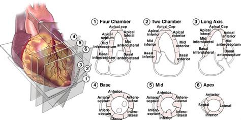
Recommendations for the Evaluation of Left Ventricular Diastolic Function by Echocardiography: An Update from the American Society of Echocardiography and the .The left ventricle is divided into 17 segments for 2D echocardiography. One can identify these segments in multiple views. The basal part is divided into six segments of 60° each. The segments along the circumference are basal anterior, basal anteroseptal, basal inferoseptal, basal inferior, basal inferolateral, and basal anterolateral.Although certain variability exists in the coronary artery blood supply to myocardial segments, segments are usually attributed to the three major coronary arteries. Visual Assessment Semi quantitative wall motion score (1-4) can be assigned to each segment to calculate the LV wall motion
Herein we review the conventional assessment of LV systolic function and examine the role of speckle-tracking echocardiography (STE), a new method to assess LV function. We also highlight the role of STE in the assessment and management of cardiac and noncardiac disease, including detection of subclinical LV dysfunction.tricular [LV] size and ejection fraction [EF], left atrial [LA] volume), outcomes data are lacking for many other parameters. Unfortunately, this approach also has limitations.Complete review on left ventricular systolic function, with emphasis on echocardiography, definitions, methods and guidelines.
A normal LV ejection fraction in the presence of the heart failure syndrome leads to a search for diastolic dysfunction. Typical echo findings in diastolic dysfunction are normal LV cavity size, thickened ventricle, and reversed E/A ratio.
LV wall has been divided into 17 segments for the ease of the assessment of regional function. Apical segment, also called apical cap, is the only segment without a direct relation to the LV cavity. These segments correspond to the three main branches of coronary blood supply as well.
Strain echocardiography, performed by using the speckle-tracking technique, can identify subclinical left ventricular dysfunction before left ventricular ejection fraction declines.This chapter demonstrates left chamber quantification through various measurements of left ventricular size and dimensions, left ventricular mass, left ventricularglobal function, regional wall motion, left ventricular segmentation, global left ventricular remodelling, and left atrial measurements.Recommendations for the Evaluation of Left Ventricular Diastolic Function by Echocardiography: An Update from the American Society of Echocardiography and the European Association of Cardiovascular Imaging.The left ventricle is divided into 17 segments for 2D echocardiography. One can identify these segments in multiple views. The basal part is divided into six segments of 60° each. The segments along the circumference are basal anterior, basal anteroseptal, basal inferoseptal, basal inferior, basal inferolateral, and basal anterolateral.
lv segments on echo lv segments diagramAlthough certain variability exists in the coronary artery blood supply to myocardial segments, segments are usually attributed to the three major coronary arteries. Visual Assessment Semi quantitative wall motion score (1-4) can be assigned to each segment to calculate the LV wall motionHerein we review the conventional assessment of LV systolic function and examine the role of speckle-tracking echocardiography (STE), a new method to assess LV function. We also highlight the role of STE in the assessment and management of cardiac and noncardiac disease, including detection of subclinical LV dysfunction.
tricular [LV] size and ejection fraction [EF], left atrial [LA] volume), outcomes data are lacking for many other parameters. Unfortunately, this approach also has limitations.
Complete review on left ventricular systolic function, with emphasis on echocardiography, definitions, methods and guidelines.
A normal LV ejection fraction in the presence of the heart failure syndrome leads to a search for diastolic dysfunction. Typical echo findings in diastolic dysfunction are normal LV cavity size, thickened ventricle, and reversed E/A ratio.
LV wall has been divided into 17 segments for the ease of the assessment of regional function. Apical segment, also called apical cap, is the only segment without a direct relation to the LV cavity. These segments correspond to the three main branches of coronary blood supply as well.
Strain echocardiography, performed by using the speckle-tracking technique, can identify subclinical left ventricular dysfunction before left ventricular ejection fraction declines.
Jun. 1. Unit. Price. Sq. Ft. Available. 4009. $2,525. 1,766 Sq. Ft. 6/1/24. New. Rockridge.
lv segments on echo|lv segments diagram